
|
BMe Research Grant |

|


Doctoral School of Physical Science
Department of Atomic Physics/Institute of Physics
Supervisor: Dr. Emőke Lőrincz
Development of PET detector module
Introducing the research area
One of today's most dynamically developing interdisciplinary area is medical physics, and in particular medical imaging, which can be divided into the following basic methods: topographic and functional imaging. One of the most advanced functional imaging techniques is Positron Emission Tomography (PET), which is widely used in cancer research, diagnosis, and medicine development.
During my PhD I participate in the development of a PET
device, more closely, working on the improvement of the image quality and spatial resolution
of PET detectors, which is an intensively researched issue in the
international literature.
Brief introduction of the research place
I conduct my PhD work in the Optical Laboratory of the Department of Atomic Physics. The department has an extensive research activity in optics-related disciplines, including basic and applied research. Our collaborations with industrial partners typically aim at the development of an equipment. Since 2006, we have been cooperating with Mediso Ltd, a developer and manufacturer of medical imaging systems. The department's research group is primarily involved in the optical design and construction, and contributed to the development of a PET device intended for human application. Results of these researches have been summarized in numerous dissertations, theses and Students' Scholarly Circles (TDK) papers.
History and context of the research
Positron Emission Tomography is a medical imaging technique that is capable of determining the location of metabolically active cells in the body. During the examination, biologically active molecules marked with a short half-life positron-emitting radiotracer are injected into the patient's body. The radiotracer (usually an analogue of glucose, marked with 12F) concentrates in the body where the uptake of that molecule is large. The patient laying on a bed is slid into the cylindrical PET device, whose inner superficies is covered with detector modules capable of sensing nuclear radiation. Upon the radioactive decay inside the body, the positron annihilates and the two radioactive particles (gamma-photons) start from the position of the decay at an angle of nearly 180 degrees between them. The detectors surrounding the patient absorb these photons, and the line at which the decay occurred is determined from these absorption positions. Image reconstruction is achieved from the data on these lines obtained during the examination using computer algorithms.
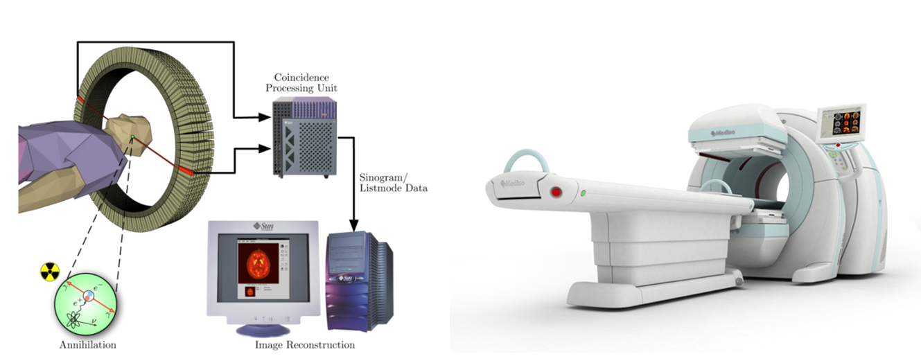
Schematic view of a PET scanner, and
the developed PET equipment
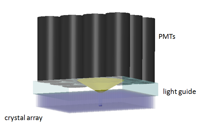
The spatial detection of the radiation is carried out with scintillator arrays comprising about 1000 pieces of small (typically 4x4x20 mm3) crystals
needles. Following the absorption of the radiation, the crystals relax from the
excited state by emitting UV/visible light; this is the so called scintillation
process. The photons emitted by the crystal are detected with photodetectors,
typically photomultiplier tubes
(PMTs). Because of the high price of the PMTs, the modules contain much less
photodetectors than crystals. Therefore, to assure the required spatial resolution a
light guide layer is placed between the crystal and the PMTs, which distributes the light
coming from the crystal among several detectors. Based on the number of
photons detected by the individual PMTs, one can determine the position of the
crystal where the scintillation occurred [1]–[5].
The research goal, open questions
The aim of my work is to improve the image quality and the spatial resolution of PET detector modules. I primarily do the experimental and simulation work related to the topic.
One drawback of the previously outlined light sharing
technique is that there are crystals (typically those located at the edges of the module) that cannot
be distinguished from each other based on the detector signals. It means that we
cannot identify the crystal where the gamma-photon was absorbed, thereby the
quality of the reconstructed image deteriorates. The solutions described in the literature typically suggest a refined calculation algorithm, which, in most
cases, does not give a satisfactory result. My goal was to
develop an optical method of modifying the
distribution of photons originating from the same scintillation on the individual
photodetectors. When optimally modifying the light distribution for the certain
crystal positions, the previously unresolvable crystals become
distinguishable.
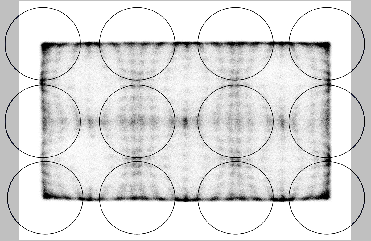
Our aim is to separate the unresolvable crystals.
Since not only the PMTs but also the crystals are quite expensive, an experimentally verified computer model was necessary to reduce technological costs. The model is expected to reliably simulate the whole detection – particularly the optical – processes.
As the optical properties of scintillator material
highly influence the spatial resolution of the detector module; acquiring precise knowledge on and experimental determination of these parameters, as well as
their integration into the computer model are important objectives of my
work.
Methodology and results
Creating a reliable computer model
The development and optimization of such a complex system
is usually done empirically, since the description of the detection process
itself requires the combination of different disciplines, i.e. nuclear physics,
optics and optoelectronics. To create a model which includes as many
physical effects as possible, I also had to investigate the underlying physical
phenomena theoretically and experimentally.
The computer model required for the detector module development was built using both of the following simulation
tools. The first program [6] is widely used in the literature but has never been validated experimentally before. The other one is an optical design tool
validated by our department’s research group previously [S1],[S2]. Both models
use Monte Carlo methods
to simulate light spread in the module, i.e. the optical effects (absorption,
reflection, etc.) are considered stochastically.
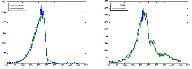
To verify the model, I carried out measurements on simple
configurations consisting of one crystal and one PMT. Combining the results of
measurements and previously validated model, I was able to identify the
critical optical parameters whose accurate examination and incorporation into
the model is necessary. Using them, the simulations were validated
experimentally [S3]–[S6].
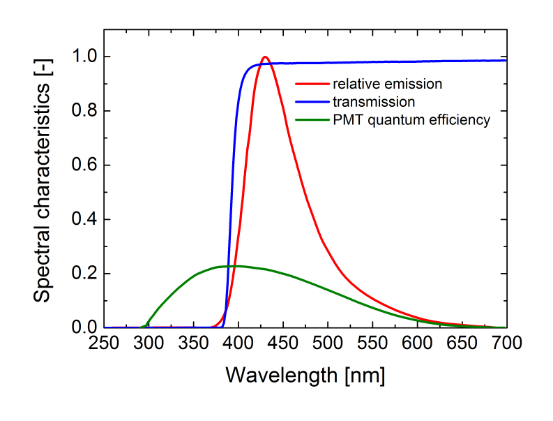
Important spectral characteristics
Improving the spatial resolution
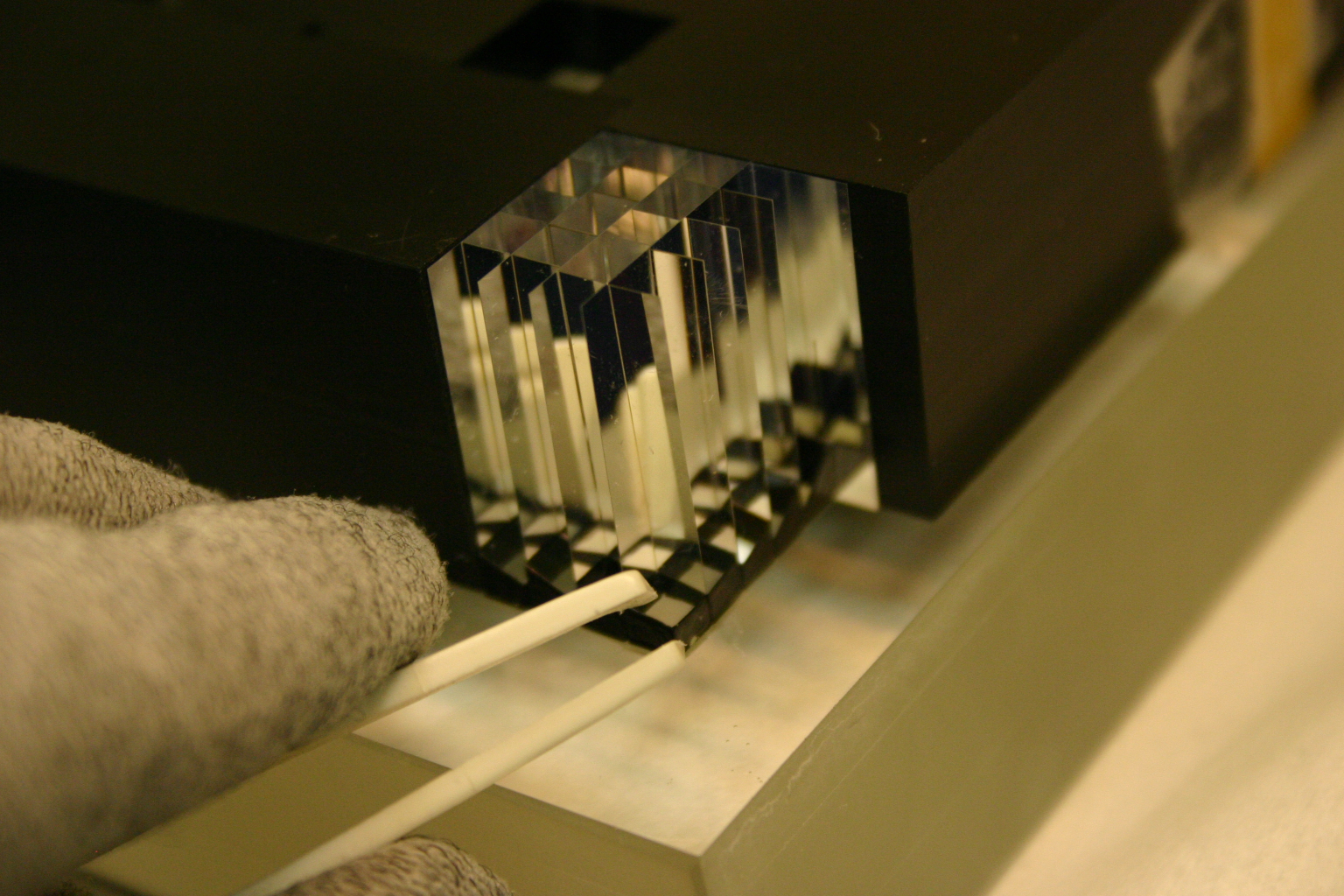
To increase the spatial resolution, we proposed a structured light guide, with the following operating principles: between the light guide and the crystals a unit should be inserted consisting of two prisms, which are separated by a small air gap. Photons arriving at the air gap with an incident angle greater than the critical angle of total internal reflection are reflected, so that only photons inside a (light)cone can spread in the second element of the double prism unit. The axis of this light cone can be modified by altering the slope angle of the air gap, thus allowing us to modify the light distribution between the photodetectors. Placing double prism units with different slope angles on adjacent, earlier indistinguishable crystals, their position can be resolved [S7].
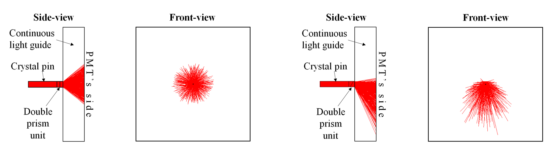
To design the special light guide, firstly I simulated the effect of the double prism unit on a single crystal, and in parallel, the same geometry was investigated experimentally. I fitted double prisms with different slope angles to the crystal, then optically coupled the crystal+prisms unit to the light guide, and analyzed the emerging light distribution. I created a computer algorithm to evaluate the measurement and compare it with the model. Taking advantage of the good agreement between the simulations and the experiment, I was able to design an optimized light guide that increases the resolution of the PET detector module.
through the cross-section profile of the light distribution
Based on the simulations, I built an experimental module consisting of crystals with optimized double prisms. With this module, which is the same as applied in the PET equipment, we are able to verify the method. Measurement on the module is in progress. At the same time, we are working on altering the special light guide so that it can be produced more easily.
Building of the experimental module
Investigation of light scattering in the crystals
The accuracy of the simulations is affected by the optical
properties of the elements of detector module (i.e. the crystals and the
photodetectors). These properties are often imperfect in the literature. I
specifically focus on the determination on the elastic light scattering within the
crystal, which slightly influences the image quality of the current detector
module. An even more significant effect occurs in case of the detector
geometry applied in PET devices that are combined with Magnetic Resonance Imaging (MRI)
scanner [7]. In the literature, light scattering was only considered based on
estimations so far. I developed a measurement method that allows us to estimate
more accurately and even to directly measure the scattering properties of the
crystals [S8].
The mechanical and optical elements required in our experiments and for the prototype of the special light guide were fabricated in the
Mechanical and Optical Workshop of our department.
Expected impact and further research
The optical technique developed in the course of my PhD work increases the PET detector module resolution, and in this way the image quality of this expensive equipment can be improved cost-effectively. The verified model is based on the investigation of the underlying physical phenomena of the detection process, and will contribute to the further developments.
The future is multimodality imaging, where
the functional and structural imaging are implemented within the same equipment.
In recent years, considerable efforts have been made to combine PET with MRI. My
measurements help take the effect of scattering into account in these
devices, while the computer model can also be utilized later when
designing a PET/MRI module.
Publications, references, links
List of publications
[S1] E. Lőrincz, G. Erdei, I. Péczeli, C. Steinbach, F. Ujhelyi, T. Bükki, I. Müller. Light Output Analyzes of Scintillator Crystal Pins and Array for PET Detector Modules. IEEE Nuclear Science Symposium and Medical Imaging Conference, pp. 4868–4871, Dresden, 2008.
[S2] E. Lőrincz, G. Erdei, I. Péczeli, C. Steinbach, F. Ujhelyi, T. Bükki, I. Müller. Modeling and optimization of scintillator array for PET detectors. IEEE Transactions on Nuclear Science, Vol. 57, No. 1, pp. 48–54, February 2010.
[S3] Steinbach C., Erdei G., Péczeli I., Ujhelyi F., Lőrincz E., Bükki T. Measuring and modelling the light efficacy of scintillator crystals for PET detector modules (in Hungarian). Kvantumelektronika, P-5, 2008.
[S4] Á. Szlávecz, B. Benyó, C. Steinbach, T. Bükki. A novel model and an environment for PET detector block simulation. Proceedings of the 7th IFAC Symposium on Modelling and Control in Biomedical Systems, Aalborg, Denmark, August 12–14, pp. 304–308, 2009.
[S5] C. O. Steinbach, Á. Szlávecz, B. Benyó, T. Bükki, and E. Lőrincz. Validation of Detect2000 based PetDetSim by simulated and measured light output of scintillator crystal pins for PET detectors. IEEE Transactions on Nuclear Science, Vol. 57, No. 5, pp. 2460–2467, October 2010.
[S6] Á. Szlávecz, T. Bükki, C. Steinbach and
B. Benyó. A novel model-based PET detector block simulation approach.
Biomedical Signal Processing and Control, Vol. 6, No. 1, pp. 27–33,
2011.
[S7] C. O. Steinbach, G. Erdei, I. Péczeli, F. Ujhelyi, T. Bükki and E. Lőrincz. Optimized Light Sharing Module for PET Block Detectors. IEEE Nuclear Science Symposium and Medical Imaging Conference, Conference Record, pp. 2817–2820, 2009.
[S8] C. O. Steinbach, F. Ujhelyi and E.
Lőrincz. Optical scattering length of LYSO scintillator crystals. IEEE Nuclear
Science Symposium and Medical Imaging Conference, 2011.
Links
BUTE Department of Atomic Physics
References
[1] M.E. Casey and R. Nutt. A Multicrystal Two Dimensional BGO Detector System for Positron Emission Tomography. IEEE Trans. Nucl. Sci., vol. 33, no. 1, pp. 460–463, February 1986.
[2] J.G. Rogers, A.J. Taylor, M.F.
Rahimi, R. Nutt, M. Andreaco, C.W. William. An Improved Multicrystal 2-D BGO
Detector for PET. IEEE Trans. Nucl. Sci., vol. 39, no. 4, pp. 1063–1068,
1992.
[3] S. Surti, J.S. Karp, R. Freifelder, E
Liu. Optimizing the Performance of a PET Detector using Discrete GSO Crystals
on a Continuous Light guide. IEEE Trans. Nucl. Sci., vol. 47, no. 3, pp.
1030–1036, June 2000.
[4] W-H. Wong, J. Uribe, K. Hicks, G. Hu. An
Analog Decoding BGO Block Detector Using Circular Photomultipliers. IEEE Trans.
Nucl. Sci., vol. 42, no. 4, pp. 1095–1101, August 1995.
[5] J.S. Karp. Against. European Journal of Nuclear Medicine, vol. 29, no. 11, pp. 1525–1528, November 2002.
[6] F. Cayouette, D. Laurendeau and C.
Moisan. DETECT2000: an improved Monte-Carlo simulator for the computer aided
design of photon sensing devices. Proc. SPIE 4833, 69 (2003)
[7] A. Ros, C.W. Lerche, F. Sanchez, J.
Segura-Ruiz, R. Ortuno, J. Marti, A. Cantarero, A. Sebastia, and J.M. Benlloch. Impact of the scattering coefficient of scintillation crystals (LYSO and LSO)
on depth of interaction resolution. IEEE Nucl. Sci. Symp. Conference Record,
2008, pp. 3715–3718.